MRI
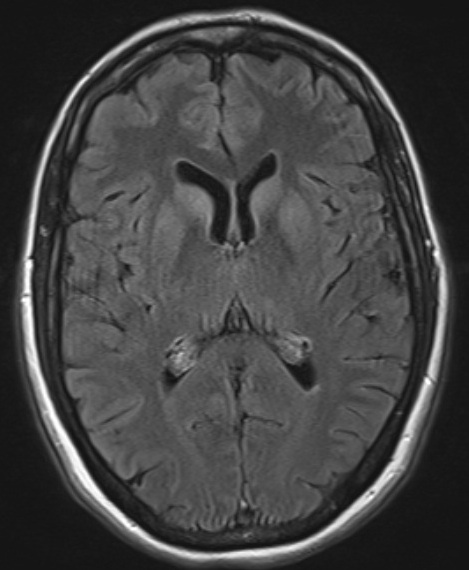
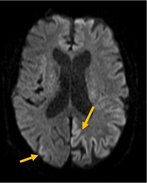
MRI is an important imaging technique used in the diagnosis of rapidly progressive dementia. In sporadic CJD hyperintensities are observed in parts of the basal ganglia the caudate nucleus, putamen, thalamus and/or strictly cortical (temporal, parietal, occipital) areas („cortical ribboning“). No subcortical white matter involvement, no isolated restricted diffusion in the thalamus. Characteristic hyperintensities may be seen on fluid attenuated inversion recovery (FLAIR) images, but diffusion weighted (DWI) sequences are required to confirm CJD-typical restricted diffusion.
CJD-typical changes are tipically seen in:
DWI

FLAIR
cortical ribboning on DWI
MRI is also helpful in differentiating sporadic and varitant CJD.
Marked hyperintense signals are found in the posterior thalamus (pulvinar sign) of patients with vCJD*

*source of image: The National Creutzfeldt-Jakob Disease Surveillance Unit, Edinburgh